Journals > > Topics > Medical Optics and Biotechnology
Medical Optics and Biotechnology|10 Article(s)
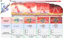
Collagen fiber anisotropy characterization by polarized photoacoustic imaging for just-in-time quantitative evaluation of burn severity
Zhenhui Zhang, Wei Chen, Dandan Cui, Jie Mi, Gen Mu, Liming Nie, Sihua Yang, and Yujiao Shi
Just-in-time burn severity assessment plays a vital role in burn treatment and care. However, it is still difficult to quantitatively and promptly evaluate burn severity by existing medical imaging methods via initial burn depth measurement since burn wounds are usually dynamically developed. As an elastic skeleton of skin, the degree of conformational changes of collagen fibers caused by overheating can reflect the burn severity in a timelier manner. Herein, the polarized photoacoustic technique (PPAT) for just-in-time quantitative evaluation of burn severity via collagen fiber anisotropy assessment is proposed. First, phantom experiments demonstrate the ability of PPAT for deep imaging in a transport mean free path and accurately quantify changes in microstructural order by thermal damage. Then, the Pearson correlation coefficient of the PPAT in assessing burn severity is shown to be up to 0.95, validated by burn skin samples. The PPAT provides a just-in-time quantitative strategy for burn severity evaluation. Just-in-time burn severity assessment plays a vital role in burn treatment and care. However, it is still difficult to quantitatively and promptly evaluate burn severity by existing medical imaging methods via initial burn depth measurement since burn wounds are usually dynamically developed. As an elastic skeleton of skin, the degree of conformational changes of collagen fibers caused by overheating can reflect the burn severity in a timelier manner. Herein, the polarized photoacoustic technique (PPAT) for just-in-time quantitative evaluation of burn severity via collagen fiber anisotropy assessment is proposed. First, phantom experiments demonstrate the ability of PPAT for deep imaging in a transport mean free path and accurately quantify changes in microstructural order by thermal damage. Then, the Pearson correlation coefficient of the PPAT in assessing burn severity is shown to be up to 0.95, validated by burn skin samples. The PPAT provides a just-in-time quantitative strategy for burn severity evaluation.
Photonics Research
- Publication Date: May. 01, 2023
- Vol. 11, Issue 5, 817 (2023)


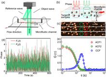
Two-beam phase correlation spectroscopy: a label-free holographic method to quantify particle flow in biofluids|On the Cover
Lan Yu, Yu Wang, Yang Wang, Kequn Zhuo, Min Liu, G. Ulrich Nienhaus, and Peng Gao
We introduce two-beam phase correlation spectroscopy (2B-ΦCS) as a label-free technique to measure the dynamics of flowing particles; e.g., in vitro or in vivo blood flow. 2B-ΦCS combines phase imaging with correlation spectroscopy, using the intrinsic refractive index contrast of particles against the fluid background in correlation analysis. This method starts with the acquisition of a time series of phase images of flowing particles using partially coherent point-diffraction digital holographic microscopy. Then, phase fluctuations from two selected circular regions in the image series are correlated to determine the concentration and flow velocity of the particles by fitting pair correlation curves with a physical model. 2B-ΦCS is a facile procedure when using a microfluidic channel, as shown by the measurements on flowing yeast microparticles, polymethyl methacrylate microparticles, and diluted rat blood. In the latter experiment, the concentration and average diameter of rat blood cells were determined to be (4.7±1.9)×106 μL-1 and 4.6±0.4 μm, respectively. We further analyzed the flow of mainly red blood cells in the tail vessels of live zebrafish embryos. Arterial and venous flow velocities were measured as 290±110 μm s-1 and 120±50 μm s-1, respectively. We envision that our technique will find applications in imaging transparent organisms and other areas of the life sciences and biomedicine. We introduce two-beam phase correlation spectroscopy (2B-ΦCS) as a label-free technique to measure the dynamics of flowing particles; e.g., in vitro or in vivo blood flow. 2B-ΦCS combines phase imaging with correlation spectroscopy, using the intrinsic refractive index contrast of particles against the fluid background in correlation analysis. This method starts with the acquisition of a time series of phase images of flowing particles using partially coherent point-diffraction digital holographic microscopy. Then, phase fluctuations from two selected circular regions in the image series are correlated to determine the concentration and flow velocity of the particles by fitting pair correlation curves with a physical model. 2B-ΦCS is a facile procedure when using a microfluidic channel, as shown by the measurements on flowing yeast microparticles, polymethyl methacrylate microparticles, and diluted rat blood. In the latter experiment, the concentration and average diameter of rat blood cells were determined to be (4.7±1.9)×106 μL-1 and 4.6±0.4 μm, respectively. We further analyzed the flow of mainly red blood cells in the tail vessels of live zebrafish embryos. Arterial and venous flow velocities were measured as 290±110 μm s-1 and 120±50 μm s-1, respectively. We envision that our technique will find applications in imaging transparent organisms and other areas of the life sciences and biomedicine.
Photonics Research
- Publication Date: Apr. 28, 2023
- Vol. 11, Issue 5, 757 (2023)
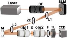
High-axial-resolution optical stimulation of neurons in vivo via two-photon optogenetics with speckle-free beaded-ring patterns
Cheng Jin, Chi Liu, and Lingjie Kong
Two-photon optogenetics has become an indispensable technology in neuroscience, due to its capability in precise and specific manipulation of neural activities. A scanless holographic approach is generally adopted to meet the requirement of stimulating neural ensembles simultaneously. However, the commonly used disk patterns fail in achieving single-neuron resolution, especially in axial dimension, and their inherent speckles decrease stimulation efficiency. Here, we propose a novel speckle-free, beaded-ring pattern for high-axial-resolution optical stimulation of neurons in vivo. Using a dye pool and a fluorescent thin film as samples, we verify that, compared to those with disk patterns, higher axial resolution and better localization ability can be achieved with beaded-ring patterns. Furthermore, we perform two-photon based all-optical physiology with neurons in mouse S1 cortex in vivo, and demonstrate that the axial resolution obtained by beaded-ring patterns can be improved by 24% when stimulating multiple neurons, compared to that of disk patterns. Two-photon optogenetics has become an indispensable technology in neuroscience, due to its capability in precise and specific manipulation of neural activities. A scanless holographic approach is generally adopted to meet the requirement of stimulating neural ensembles simultaneously. However, the commonly used disk patterns fail in achieving single-neuron resolution, especially in axial dimension, and their inherent speckles decrease stimulation efficiency. Here, we propose a novel speckle-free, beaded-ring pattern for high-axial-resolution optical stimulation of neurons in vivo. Using a dye pool and a fluorescent thin film as samples, we verify that, compared to those with disk patterns, higher axial resolution and better localization ability can be achieved with beaded-ring patterns. Furthermore, we perform two-photon based all-optical physiology with neurons in mouse S1 cortex in vivo, and demonstrate that the axial resolution obtained by beaded-ring patterns can be improved by 24% when stimulating multiple neurons, compared to that of disk patterns.
Photonics Research
- Publication Date: May. 12, 2022
- Vol. 10, Issue 6, 06001367 (2022)
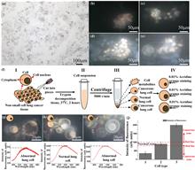
Single-cell detection by enhancement of fluorescence in waveguides for cancer diagnosis and therapy
Hailang Dai, Hongrui Shan, Zhangchi Sun, Daopeng Dai, Yuxi Shang, Zhuangqi Cao, and Xianfeng Chen
Cancer is one of the most common diseases to threaten human health. If individuals are diagnosed with malignant tumors via a single cell, medical workers are greatly advantageous to early diagnose and intervene in malignant tumors therapy. In this paper, we propose a fluorescence detection map to rapidly distinguish whether the chromosomes of a cell are normal or abnormal by detecting the fluorescent intensity of a single cell. Herein, we draw a map from a single cell with an abnormal number of chromosomes that is monitored in real time. Moreover, this way offers precise and prompt detection of the surviving of cancer cells at or near the site of the tumor after treatments for cancer, which can achieve personalized cancer diagnosis and therapy. Therefore, cancer recurrences and metastasis can be effectively identified, utilizing this ultrasensitive detection method of an abnormal chromosome number. Cancer is one of the most common diseases to threaten human health. If individuals are diagnosed with malignant tumors via a single cell, medical workers are greatly advantageous to early diagnose and intervene in malignant tumors therapy. In this paper, we propose a fluorescence detection map to rapidly distinguish whether the chromosomes of a cell are normal or abnormal by detecting the fluorescent intensity of a single cell. Herein, we draw a map from a single cell with an abnormal number of chromosomes that is monitored in real time. Moreover, this way offers precise and prompt detection of the surviving of cancer cells at or near the site of the tumor after treatments for cancer, which can achieve personalized cancer diagnosis and therapy. Therefore, cancer recurrences and metastasis can be effectively identified, utilizing this ultrasensitive detection method of an abnormal chromosome number.
Photonics Research
- Publication Date: Nov. 15, 2021
- Vol. 9, Issue 12, 12002381 (2021)
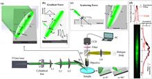
Distance-controllable and direction-steerable opto-conveyor for targeting delivery
Zhen Che, Wenguo Zhu, Yaoming Huang, Yu Zhang, Linqing Zhuo, Pengpeng Fan, Zhibin Li, Huadan Zheng, Wenjin Long, Wentao Qiu, Yunhan Luo, Jun Zhang, Jinghua Ge, Jianhui Yu, and Zhe Chen
Opto-conveyors have attracted widespread interest in various fields because of their non-invasive and non-contact delivery of micro/nanoparticles. However, the flexible control of the delivery distance and the dynamic steering of the delivery direction, although very desirable in all-optical manipulation, have not yet been achieved by opto-conveyors. Here, using a simple and cost-effective scheme of an elliptically focused laser beam obliquely irradiated on a substrate, a direction-steerable and distance-controllable opto-conveyor for the targeting delivery of microparticles is implemented. Theoretically, in the proposed scheme of the opto-conveyor, the transverse and longitudinal resultant forces of the optical gradient force and the optical scattering force result in the transverse confinement and the longitudinal transportation of microparticles, respectively. In this study, it is experimentally shown that the proposed opto-conveyor is capable of realizing the targeting delivery for microparticles. Additionally, the delivery distance of microparticles can be flexibly and precisely controlled by simply adjusting the irradiation time. By simply rotating the cylindrical lens, the proposed opto-conveyor is capable of steering the delivery direction flexibly within a large range of azimuthal angles, from ?75° to 75°. This study also successfully demonstrated the real-time dynamic steering of the delivery direction from ?45° to 45° with the dynamical rotation of the cylindrical lens. Owing to its simplicity, flexibility, and controllability, the proposed method is capable of creating new opportunities in bioassays as well as in drug delivery. Opto-conveyors have attracted widespread interest in various fields because of their non-invasive and non-contact delivery of micro/nanoparticles. However, the flexible control of the delivery distance and the dynamic steering of the delivery direction, although very desirable in all-optical manipulation, have not yet been achieved by opto-conveyors. Here, using a simple and cost-effective scheme of an elliptically focused laser beam obliquely irradiated on a substrate, a direction-steerable and distance-controllable opto-conveyor for the targeting delivery of microparticles is implemented. Theoretically, in the proposed scheme of the opto-conveyor, the transverse and longitudinal resultant forces of the optical gradient force and the optical scattering force result in the transverse confinement and the longitudinal transportation of microparticles, respectively. In this study, it is experimentally shown that the proposed opto-conveyor is capable of realizing the targeting delivery for microparticles. Additionally, the delivery distance of microparticles can be flexibly and precisely controlled by simply adjusting the irradiation time. By simply rotating the cylindrical lens, the proposed opto-conveyor is capable of steering the delivery direction flexibly within a large range of azimuthal angles, from ?75° to 75°. This study also successfully demonstrated the real-time dynamic steering of the delivery direction from ?45° to 45° with the dynamical rotation of the cylindrical lens. Owing to its simplicity, flexibility, and controllability, the proposed method is capable of creating new opportunities in bioassays as well as in drug delivery.
Photonics Research
- Publication Date: Jun. 05, 2020
- Vol. 8, Issue 7, 07001124 (2020)

Stimulated Raman scattering signal generation in a scattering medium using self-reconstructing Bessel beams
Xueli Chen, Xinyu Wang, Lin Wang, Peng Lin, Yonghua Zhan, and Ji-Xin Cheng
Scattering is a huge challenge for microscopic imaging. Indeed, it is difficult to observe target chemicals in scattering media by means of the current Gaussian beam-based stimulated Raman scattering (SRS) microscopy, since the tight focus of the Gaussian beam is destroyed after propagating through a certain distance. Bessel beams, featuring self-reconstructing property, may bring a solution to this problem. By combining Bessel beams with SRS microscopy, we can probe the SRS signal from a scattering medium. In this paper, using the beam propagation method, we first simulate the propagation of the Bessel beam as well as the generation and self-reconstruction of SRS signals. By adding glass beads along the beam propagation path in order to simulate scattering, the propagation of the Bessel beams and the generation of the SRS signals will change. Then, we design a series of simulations to investigate the influence of the size, position, number, and distribution of the added glass beads on the generation of the SRS signals. A preliminary experiment is also carried out to confirm the simulation predictions. Results demonstrate that the SRS signals can be generated or be recovered at a certain depth in scattering media, and that such signals are greatly affected by the parameters of the scatters. Scattering is a huge challenge for microscopic imaging. Indeed, it is difficult to observe target chemicals in scattering media by means of the current Gaussian beam-based stimulated Raman scattering (SRS) microscopy, since the tight focus of the Gaussian beam is destroyed after propagating through a certain distance. Bessel beams, featuring self-reconstructing property, may bring a solution to this problem. By combining Bessel beams with SRS microscopy, we can probe the SRS signal from a scattering medium. In this paper, using the beam propagation method, we first simulate the propagation of the Bessel beam as well as the generation and self-reconstruction of SRS signals. By adding glass beads along the beam propagation path in order to simulate scattering, the propagation of the Bessel beams and the generation of the SRS signals will change. Then, we design a series of simulations to investigate the influence of the size, position, number, and distribution of the added glass beads on the generation of the SRS signals. A preliminary experiment is also carried out to confirm the simulation predictions. Results demonstrate that the SRS signals can be generated or be recovered at a certain depth in scattering media, and that such signals are greatly affected by the parameters of the scatters.
Photonics Research
- Publication Date: May. 26, 2020
- Vol. 8, Issue 6, 06000929 (2020)
Dark mode plasmonic optical microcavity biochemical sensor
Cheng Li, Lei Chen, Euan McLeod, and Judith Su
Whispering gallery mode (WGM) microtoroid optical resonators have been effectively used to sense low concentrations of biomolecules down to the single molecule limit. Optical WGM biochemical sensors such as the microtoroid operate by tracking changes in resonant frequency as particles enter the evanescent near field of the resonator. Previously, gold nanoparticles have been coupled to WGM resonators to increase the magnitude of resonance shifts via plasmonic enhancement of the electric field. However, this approach results in increased scattering from the WGM, which degrades its quality (Q) factor, making it less sensitive to extremely small frequency shifts caused by small molecules or protein conformational changes. Here, we show using simulation that precisely positioned trimer gold nanostructures generate dark modes that suppress radiation loss and can achieve high (>106) Q with an electric-field intensity enhancement of 4300, which far exceeds that of a single rod (~2500 times). Through an overall evaluation of a combined enhancement factor, which includes the Q factor of the system, the sensitivity of the trimer system was improved 105× versus 84× for a single rod. Further simulations demonstrate that unlike a single rod system, the trimer is robust to orientation changes and has increased capture area. We also conduct stability tests to show that small positioning errors do not greatly impact the result. Whispering gallery mode (WGM) microtoroid optical resonators have been effectively used to sense low concentrations of biomolecules down to the single molecule limit. Optical WGM biochemical sensors such as the microtoroid operate by tracking changes in resonant frequency as particles enter the evanescent near field of the resonator. Previously, gold nanoparticles have been coupled to WGM resonators to increase the magnitude of resonance shifts via plasmonic enhancement of the electric field. However, this approach results in increased scattering from the WGM, which degrades its quality (Q) factor, making it less sensitive to extremely small frequency shifts caused by small molecules or protein conformational changes. Here, we show using simulation that precisely positioned trimer gold nanostructures generate dark modes that suppress radiation loss and can achieve high (>106) Q with an electric-field intensity enhancement of 4300, which far exceeds that of a single rod (~2500 times). Through an overall evaluation of a combined enhancement factor, which includes the Q factor of the system, the sensitivity of the trimer system was improved 105× versus 84× for a single rod. Further simulations demonstrate that unlike a single rod system, the trimer is robust to orientation changes and has increased capture area. We also conduct stability tests to show that small positioning errors do not greatly impact the result.
Photonics Research
- Publication Date: Aug. 01, 2019
- Vol. 7, Issue 8, 08000939 (2019)
Physical picture of the optical memory effect|Editors' Pick
Honglin Liu, Zhentao Liu, Meijun Chen, Shensheng Han, and Lihong V. Wang
The optical memory effect is an interesting phenomenon that has attracted considerable attention in recent decades. Here, we present a new physical picture of the optical memory effect, in which the memory effect and the conventional spatial shift invariance are united. Based on this picture we depict the role of thickness, scattering times, and anisotropy factor and derive equations to calculate the ranges of the angular memory effect (AME) of different scattering components (ballistic light, singly scattered, doubly scattered, etc.), and hence a more accurate equation for the real AME ranges of volumetric turbid media. A conventional random phase mask model is modified according to the new picture. The self-consistency of the simulation model and its agreement with the experiment demonstrate the rationality of the model and the physical picture, which provide powerful tools for more sophisticated studies of the memory-effect-related phenomena and wavefront-sensitive techniques, such as wavefront shaping, optical phase conjugation, and optical trapping in/through scattering media. The optical memory effect is an interesting phenomenon that has attracted considerable attention in recent decades. Here, we present a new physical picture of the optical memory effect, in which the memory effect and the conventional spatial shift invariance are united. Based on this picture we depict the role of thickness, scattering times, and anisotropy factor and derive equations to calculate the ranges of the angular memory effect (AME) of different scattering components (ballistic light, singly scattered, doubly scattered, etc.), and hence a more accurate equation for the real AME ranges of volumetric turbid media. A conventional random phase mask model is modified according to the new picture. The self-consistency of the simulation model and its agreement with the experiment demonstrate the rationality of the model and the physical picture, which provide powerful tools for more sophisticated studies of the memory-effect-related phenomena and wavefront-sensitive techniques, such as wavefront shaping, optical phase conjugation, and optical trapping in/through scattering media.
Photonics Research
- Publication Date: Nov. 01, 2019
- Vol. 7, Issue 11, 11001323 (2019)
Irreversible denaturation of DNA: a method to precisely control the optical and thermo-optic properties of DNA thin solid films
Hayoung Jeong, Paulson Bjorn, Seongjin Hong, Seunguk Cheon, and Kyunghwan Oh
The denaturation of double-stranded deoxyribonucleic acid (ds-DNA) has been well known to break nucleobase bonds, resulting in single-stranded deoxyribonucleic acid (ss-DNA) in solutions, which can recombine to form ds-DNA in a reversible manner. We developed an efficient process to irreversibly maintain various DNA denaturation levels in thin solid films in order to investigate the impacts of the denaturation on the optical properties of DNA films. By adding NaOH in an aqueous solution of salmon testis DNA, we flexibly controlled the level of denaturation in the solution, which was then spin-coated on Si and silica substrates to irreversibly bind ss-DNAs in a thin solid film. The denaturation of DNA in thin solid films was experimentally confirmed by ultraviolet-visible and Fourier transform infrared spectroscopic investigations, whose level could be controlled by the NaOH content in the aqueous solution precursor. By this irreversible denaturation process, we developed a new method to flexibly vary the refractive index of DNA thin solid films in a wide range of Δn>0.02 in the visible to near-infrared range. Thermo-optic coefficients dn/dT of the films were also experimentally measured in the temperature range from 40°C to 90°C to confirm the significant impacts of denaturation. Detailed thin film processes and optical characterizations are discussed. The denaturation of double-stranded deoxyribonucleic acid (ds-DNA) has been well known to break nucleobase bonds, resulting in single-stranded deoxyribonucleic acid (ss-DNA) in solutions, which can recombine to form ds-DNA in a reversible manner. We developed an efficient process to irreversibly maintain various DNA denaturation levels in thin solid films in order to investigate the impacts of the denaturation on the optical properties of DNA films. By adding NaOH in an aqueous solution of salmon testis DNA, we flexibly controlled the level of denaturation in the solution, which was then spin-coated on Si and silica substrates to irreversibly bind ss-DNAs in a thin solid film. The denaturation of DNA in thin solid films was experimentally confirmed by ultraviolet-visible and Fourier transform infrared spectroscopic investigations, whose level could be controlled by the NaOH content in the aqueous solution precursor. By this irreversible denaturation process, we developed a new method to flexibly vary the refractive index of DNA thin solid films in a wide range of Δn>0.02 in the visible to near-infrared range. Thermo-optic coefficients dn/dT of the films were also experimentally measured in the temperature range from 40°C to 90°C to confirm the significant impacts of denaturation. Detailed thin film processes and optical characterizations are discussed.
Photonics Research
- Publication Date: Aug. 22, 2018
- Vol. 6, Issue 9, 09000918 (2018)
Coherent optical adaptive technique improves the spatial resolution of STED microscopy in thick samples
Wei Yan, Yanlong Yang, Yu Tan, Xun Chen, Yang Li, Junle Qu, and Tong Ye
Stimulated emission depletion (STED) microscopy is one of far-field optical microscopy techniques that can provide sub-diffraction spatial resolution. The spatial resolution of the STED microscopy is determined by the specially engineered beam profile of the depletion beam and its power. However, the beam profile of the depletion beam may be distorted due to aberrations of optical systems and inhomogeneity of a specimen’s optical properties, resulting in a compromised spatial resolution. The situation gets deteriorated when thick samples are imaged. In the worst case, the severe distortion of the depletion beam profile may cause complete loss of the super-resolution effect no matter how much depletion power is applied to specimens. Previously several adaptive optics approaches have been explored to compensate aberrations of systems and specimens. However, it is difficult to correct the complicated high-order optical aberrations of specimens. In this report, we demonstrate that the complicated distorted wavefront from a thick phantom sample can be measured by using the coherent optical adaptive technique. The full correction can effectively maintain and improve spatial resolution in imaging thick samples. Stimulated emission depletion (STED) microscopy is one of far-field optical microscopy techniques that can provide sub-diffraction spatial resolution. The spatial resolution of the STED microscopy is determined by the specially engineered beam profile of the depletion beam and its power. However, the beam profile of the depletion beam may be distorted due to aberrations of optical systems and inhomogeneity of a specimen’s optical properties, resulting in a compromised spatial resolution. The situation gets deteriorated when thick samples are imaged. In the worst case, the severe distortion of the depletion beam profile may cause complete loss of the super-resolution effect no matter how much depletion power is applied to specimens. Previously several adaptive optics approaches have been explored to compensate aberrations of systems and specimens. However, it is difficult to correct the complicated high-order optical aberrations of specimens. In this report, we demonstrate that the complicated distorted wavefront from a thick phantom sample can be measured by using the coherent optical adaptive technique. The full correction can effectively maintain and improve spatial resolution in imaging thick samples.
Photonics Research
- Publication Date: Apr. 12, 2017
- Vol. 5, Issue 3, 03000176 (2017)
Topics
© Copyright 2018-2021 | Chinese Laser Press. All Rights Reserved 沪ICP备15018463号-20